- #Free dicom viewer and image analysis how to#
- #Free dicom viewer and image analysis install#
- #Free dicom viewer and image analysis software#
- #Free dicom viewer and image analysis professional#
- #Free dicom viewer and image analysis series#
We hope that you found valuable information here to facilitate your everyday work as our main concern is about helping and forming human and animal caregivers.
#Free dicom viewer and image analysis professional#
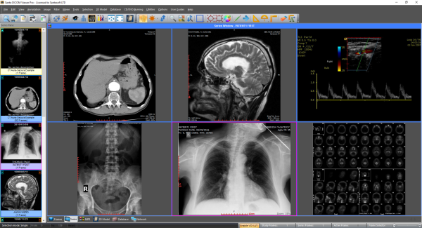
⚠ Scrolling images not available: severely restricts the professional use Finally, a multi-lingual support is available. Moreover, it supports touch screens and is easy to share. As it supports multi-windows, you can open and analyze multiple images simultaneously using it. The supported formats include TIFF, GIF, JPEG, BMP, DICOM, FITS, and raw images. It can be used to view, edit, process, and analyze 8-bit, 16-bit, and 32-bit images.
#Free dicom viewer and image analysis software#

Dicom Library supports many DICOM modalities and PACS models.

This is particularly useful when doctors are running away from their DICOM workstations and DICOM-ready computers.
#Free dicom viewer and image analysis install#
We have only selected online web-based DICOM viewers because it definitely saves time for healthcare professionals to install desktop DICOM viewers on local computers. To view these images on your computer, you will have to use a DICOM viewer, which will interpret the file information and display it as an image. Inconveniently, in contrast to other image file formats such as JPEG or TIFF, DICOM files are not recognized by Operating System(Windows®, Mac)as image files.
#Free dicom viewer and image analysis series#
The information within the header is organized as a constant and standardized series of tags. A DICOM file consists of a header and image data sets packed into a single file.
#Free dicom viewer and image analysis how to#
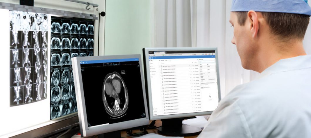
It is an international standard for medical imaging. Here i go over all the methods for viewing DICOM images on a Mac, from free to monthly fee, cloud based to disk based.Figuring out how to view a DICOM image on a Mac can be a frustrating process. What is a DICOM file or DICOM image?ĭICOM stands for “Digital Imaging and Communications in Medicine”. Several image management software packages that permit easy screen capture (such as. The software can read over 30 varieties of DICOM format, including films – Cine mode.Have you ever experienced the never-ending wait for a patient CD to open? Or wasting time trying to load data using your tablet instead of your desktop? This is definitely an avoidable loss of time if you find the DICOM viewer that corresponds to your personal use. Several free stand-alone DICOM browsers (such as DicomWorks, ImageJn and MEDISP Viewer) available on the Internet are good examples of such software.7,8,14 An earlier article in this series has reviewed the capabilities and limitations of free DICOM browsers. The browser supports medical images in the DICOM 3.0 format. As a result, the doctor can describe the study as soon as it is sent from the device to the PACS server. As the application can be made available to physicians directly from hospital servers, there is no need to send a large number of DICOM files to descriptive stations located outside the hospital. The clinical client is intended for departmental physicians so that they can review the study at a reference station located, for example, in a ward.ĭO system architecture allows for easy and definitely faster description of teleradiological tests. The diagnostic client is intended for radiologists to view the tests at a descriptive station which the corresponding diagnostic monitors are connected to. Depending on the configuration, the application can operate in two modes: diagnostic and clinical ones. It works with medical devices supporting DICOM 3.0 communication protocols. Exhibeon is a solution addressed to doctors, technicians and is used to search, view and analyze digital photos in the DICOM standard.

We give you the application for viewing and medical diagnostics of medical tests (CT, MR, X-ray, Mammography, etc.). in a much shorter time than it would be on the aforementioned workstation. This solution allows you to perform advanced 3D reconstructions: MPR, VOL, MIP, minIP, etc.

The architecture of the Image Distribution system has been designed to maximize the use of hardware resources and to reduce the waiting time for the use of the tool, therefore calculations are performed on a dedicated server, which in a minimum configuration is much stronger than the basic workstation located in the branch.


 0 kommentar(er)
